Menu
e-shop


Vlašín M., Ceplecha V. Stenotická tenosynovitida m. abductor pollicis longus u závodního saňového
psa – kazuistika. Veterinářství 2026;76(11):589-593.
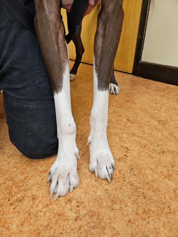
Cíl práce: Popsat diagnostický postup a konzervativní léčbu onemocnění šlachy m. abductor pollicis longus (APL) u mladého závodního saňového psa s recidivujícím zduřením v oblasti pravého karpu. Kasuistika: Nekastrovaný pes ve věku 1,5 roku byl vyšetřen pro recidivující zduření v oblasti pravého karpu. Obtíže vznikly přibližně čtyři týdny před vyšetřením po napadení jiným psem, kdy pacient začal kulhat na pravou hrudní končetinu a z poranění vytékala tekutina. Po primárním ošetření rány došlo během pěti dnů k úpravě klinického stavu. Přibližně o týden později se objevilo zduření dorzomediálně nad pravým karpem, které reagovalo na podávání nesteroidních antiflogistik. Ke dvěma relapsům došlo vždy po návratu k tréninkové zátěži. Při ortopedickém vyšetření byl pacient bez kulhání, se zachovaným rozsahem pohybu v karpu. Přítomno bylo tuhé, nefluktuující, nebolestivé zduření dorzomediálně nad pravým karpem a mírný diskomfort při maximální flexi. CT vyšetření prokázalo zesílení úponové šlachy APL a ložiskové změny v subchondrální kosti distálního radia v oblasti radiokarpálního kloubu. Ultrasonografické vyšetření zobrazilo změny šlachových vláken a edém v oblasti šlachy a šlachové pochvy. Nález byl kompatibilní s tendinopatií až stenotickou tenosynovitidou APL. Pacient byl léčen konzervativně pomocí fyzioterapie (rázové vlny, terapeutického laseru a kontrolované postupné zátěže). Dva měsíce po zahájení léčby nekulhal a postupně se vracel k plnému tréninku. Závěr: U psů s recidivujícím zduřením v oblasti karpu je vhodné zahrnout onemocnění m. abductor policis longus (APL) mezi diferenciální diagnózy. Kombinace CT a ultrasonografického vyšetření umožňuje posoudit jak změny měkkých tkání, tak případné změny přilehlých kostních struktur. U klinicky stabilního pacienta může být vhodnou první volbou konzervativní terapie s řízenou rehabilitací.
Klíčová slova: pes, karpus, m. abductor pollicis longus, tendinopatie, tenosynovitida, ultrasonografie, počítačová tomografie, sportovní pes
Objective To describe the diagnostic approach and conservative management of abductor pollicis longus (APL)tendon disease in a young racing sled dog presenting with recurrent swelling of the right carpal region.
Case description A 1.5-year-old intact male dog was evaluated for recurrent swelling of the right carpal region. Clinical signs developed approximately four weeks before presentation following an attack by another dog, after which the patient developed right thoracic limb lameness associated with fluid discharge from the wound. Following initial wound management, the clinical signs resolved within five days. Approximately one week later, swelling developed over the dorsomedial aspect of the right carpus and responded to treatment with non-steroidal anti-inflammatory drugs. Two subsequent episodes of recurrence occurred following return to training. At orthopaedic examination, the dog was not lame and had a normal range of motion of the carpus. A firm, non-fluctuant, non-painful swelling was present over the dorsomedial aspect of the right carpus, with mild discomfort elicited at maximal carpal flexion. Computed tomography (CT) demonstrated thickening of the insertional portion of the APL tendon and focal changes within the subchondral bone of the distal radius adjacent to the radiocarpal joint. Ultrasonographic examination revealed alterations in the tendon fibre architecture and oedema involving the tendon and its tendon sheath. The findings were considered consistent with APL tendinopathy to stenosing tenosynovitis. The patient was managed conservatively with physiotherapy (extracorporeal shockwave therapy, therapeutic laser treatment, and controlled, progressive return to exercise). Two months after initiation of treatment, the dog remained free of lameness and was gradually returning to full training. Conclusion :In dogs presenting with recurrent swelling of the carpal region, APL tendon disease should be included among the differential diagnoses. The combination of CT and ultrasonography allows assessment of both soft-tissue abnormalities and potential changes in adjacent osseous structures. In clinically stable patients, conservative management combined with a structured rehabilitation programme may represent an appropriate first-line treatment option.
Keywords: dog, carpus, m. abductor pollicis longus, tendinopathy, tenosynovitis, ultrasonography, computed
tomography, sporting dog