Ceplecha V., Proks P., Řeháková K. Etiopatogeneze, diagnostika a léčba jaterní fibrózy u psů. Etiopathogenesis, diagnosis and therapy of hepatic fibrosis in dogs. Veterinární klinika. 2020;17(1):33-40.
SOUHRN
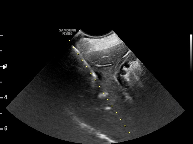
Jaterní fibróza je charakterizována jako zmnožení vaziva v jaterní tkáni. Funkční tkáň jater je tak pod vlivem progrese onemocnění postupně nahrazována nově vznikající vazivovou tkání. V konečném stadiu, kdy je jaterní parenchym kompletně přestavěn a fibrotizaci provází změny cévního řečiště i žlučovodů, přechází fibróza v cirhózu. Klíčovou roli v etiopatogenezi jaterní fibrózy sehrávají jaterní stelární buňky (Itóovy buňky), jejichž aktivací dochází k fibrogenezi, tedy produkci extracelulární matrix. Přebytek extracelulární matrix však indukuje fibrolýzu, tedy degradaci vaziva. Fibróza je proto považována za dynamický proces a na rozdíl od cirhózy také za reverzibilní. Z toho důvodu je kladen důraz na časnou diagnostiku jaterního onemocnění s následnou specifickou kauzální a antifibrotickou terapií. K fibróze jater vede řada hepatobiliárních onemocnění – např. chronická hepatitida, extrahepatální obstrukce žlučových cest nebo vrozené abnormality duktální ploténky. Pokročilá fibróza, resp. cirhóza se mohou projevit hepatální encefalopatií, koagulopatiemi, ikterem a příznaky portální hypertenze. Rutinní ultrasonografické vyšetření často není dostatečně senzitivní pro odlišení zdravé jaterní tkáně od tkáně patologické. Zlatým standardem diagnostiky tak zůstává histopatologické vyšetření jaterního bioptátu, které nám v mnoha případech rovněž umožní rozpoznat příčinu jaterní fibrózy. Z neinvazivních markerů fibrózy jater lze zmínit slibné výsledky studií, které se zabývaly stanovením kys. hyaluronové nebo poměru AST k počtu trombocytů.
SUMMARY
Hepatic fibrosis is characterized by multiplication of fibrous tissue in the liver tissue. Thus, functional liver tissue is under the influence of disease progression gradually substituted newly arising fibrous tissue. In final stage, when the liver parenchyma is completely rebuilt and fibrosing is accompanied by changes in the vascular bed and bile ducts, fibrosis is rebuilt in cirrhosis. Key role in the etiopathogenesis of hepatic fibrosis play liver stellate cells (Ito cells), which activation results in fibrogenesis, i.e. a production of the extracellular matrix. However, an excess of the extracellular matrix induces fibrolysis, i.e. degradation of the fibrous tissue. Fibrosis is therefore considered a dynamic process and, unlike cirrhosis, also reversible. Therefore, emphasis is placed on early diagnosis of the liver disease followed by specific causal and anti-fibrous treatment. A number of hepatobiliary diseases lead to hepatic fibrosis, for example chronic hepatitis, extrahepatic biliary ducts obstruction or congenital abnormalities of the ductal plate. Advanced fibrosis, resp. cirrhosis may present as hepatic encephalopathy, coagulopathies, icterus and signs of the portal hypertension. Routine ultrasonographic examination is often not sensitive enough for differentiation of healthy hepatic tissue from pathological tissue. The histopathological examination of liver biopsy remains the gold standard of diagnosis that in many cases allows recognize the cause of hepatic fibrosis. Among the non-invasive markers of hepatic fibrosis, the promising results of studies on the determination of hyaluronic acid or the ratio of AST to platelet counts can be mentioned.*